Dental Imaging
As you are likely well aware, dental x-rays provide your dentist with extremely important information they need in order to better help you care for your teeth, preventing decayed and injured teeth where possible and restoring decayed and injured teeth where necessary. These x-rays allow your dentist to see below the surfaces that are visible in your mouth and even detect anomalies that are just beginning to arise so that they can be handled. However, there are some cases where traditional x-rays are insufficient in providing your dentist with the full information they need for efficient treatment planning, and another form of more thorough dental imaging must be used.
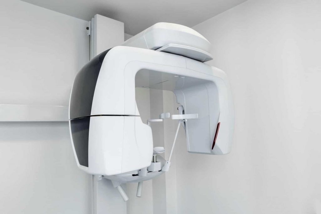
Dental Cone Beam Computed Tomography
Dental cone beam computed tomography, or CT, is not used as frequently as traditional dental x-rays, but they can be essential in cases where your dentist needs to see what is occurring beneath the surface of your teeth and gums eve more clearly than traditional x-rays allow. A cone-shaped x-ray beam is moved around the patient’s head in order to produce a large number of high-quality views. The technology that CT scanners use allow for the generation of three dimensional images of dental structure, soft tissue, nerve paths and bone in just one scan. These images can allow the dentist to evaluate many conditions that are related to oral and overall health, including diseases that occur in the jaw, dentition, bony facial structures, nasal cavity and sinuses, so as to better address and resolve these issues. It can also help in cases where:
- The patient has impacted teeth and surgery must be planned.
- The patient may have temporomandibular joint disorder (more commonly referred to as TMJ) and needs a formal diagnosis.
- The patient needs dental implants which require very precise placement.
- The dentist needs to perform a thorough evaluation of the jaw, sinuses, nerve canals and nasal cavity for either preventative or restorative purposes.
- The dentist suspects that the patient may have jaw tumors that need to be accurately detected, measured and treated.
- The patient is suffering from pain or disease and the origin needs to be determined.
- The patient will benefit from cephalometric analysis.
- The dentist needs to determine bone structure and tooth orientation.
- The patient needs reconstructive surgery.
Receiving Dental Cone Beam CT
Your dentist will recommend that you wear comfortable, loose-fitting clothing if you are going to have a dental CT scan performed. You should also avoid wearing any metal objects, including jewelry and hairpins, as these can adversely affect the images produced. In some cases your dentist may also recommend that you remove eyeglasses, piercings, hearing aids and any removable dental work.
Depending on the scanner and the area of particular focus, you will either sit or lie down for the dental cone beam CT scan. The scanner gantry will rotate around your head a complete 360 degrees in order to produce the many views that are necessary to form a detailed three-dimensional image. It is very important that you remain as still as possible throughout the scan, which can take anywhere between ten to forty seconds.
A dental cone beam CT produces high-quality images that present the many detailed angles your dentist needs in order to better evaluate, diagnose and treat conditions relating to your oral health. It is painless, noninvasive, and far more accurate than traditional dental x-rays, with a unique ability to image both bone and soft tissue at the same time. In many cases, this can make dental cone beam CT far more beneficial, especially where a patient is suffering from oral health issues that require aggressive attention.
For more information about this specialized dental imaging and whether it’s right for you, contact Dr, Nurminsky today.
